Digital Fluorescein Angiography and Fundus Photography
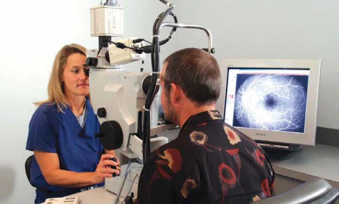
A large digital camera, a computer server, and a color monitor are the essential components of a digital photography and angiography system. High resolution photographs are taken of the patient's retina by a trained retinal photographer, and the results are available immediately for review by the doctor.
Digital retinal photography is performed with a large digital camera that takes high resolution photographs of the retina and macula. The test is performed with the patient seated in a chair, and both the camera and photographer facing the patient. The images are available immediately for review on a computer monitor by both the doctor and the patient. Digital retinal photography has many clinical uses. For example, it is particularly useful for accurately documenting the extent of retinal disease at one point in time, for comparison at a later date.
Fluorescein (a yellow synthetic dye) angiography may also be used to enhance the digital photographs in certain retinal diseases. Fluorescein is injected into a vein in the arm and circulates quickly to the eye. When the digital camera is used with certain light filters, fluorescein will highlight blood vessels and most areas of retinal disease. Clinical decision making for a variety of retinal diseases depends on fluorescein angiography. For example, fluorescein angiography is commonly used to guide treatment for age-related macular degeneration, diabetic retinopathy, and retinal vascular occlusions.
In the past, 35 mm film was used for retinal photography and angiography. This requires waiting for the film to be developed for a clinical decision to be made. This can take several days if the film is sent off-site for development. With the latest technology, digital angiography and photography provide instant and accurate results viewable on a computer monitor or by hard copy from a photographic printer.