Retinal Tears & Retinal Detachment

Retinal tears and detachments are sight-threatening conditions that are considered one of the few ocular emergencies. A retinal detachment can occur at any age, but it is more common in people over 40 years of age. It affects men more than women. It is estimated that 1 in 300 people develop a retinal detachment sometime during their life.
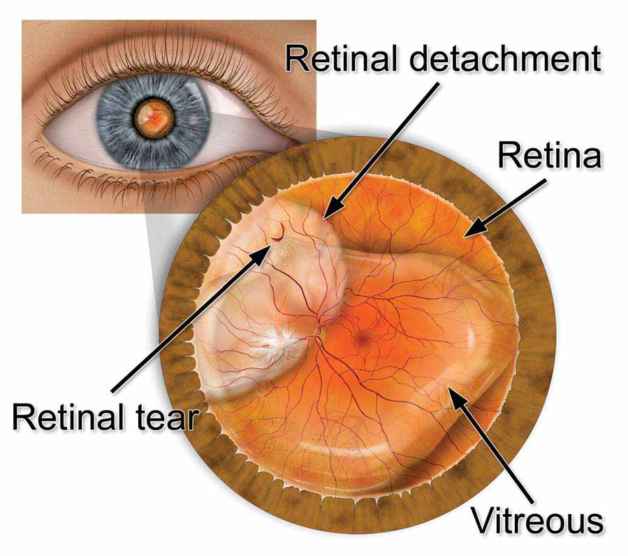
The retina is a thin layer of nerve tissue that lines the inside of the eye. This nerve layer is analogous to the "film inside a camera" and is essential for sight. In certain abnormal conditions, the retina may separate from the inside wall of the eye and hang freely within the middle of the eye. This is called a retinal detachment.
The most common sequence of events leading to a retinal detachment begins with the sudden collapse of the vitreous gel that fills the middle of the eye, called a posterior vitreous detachment (PVD). As the vitreous collapses, it may pull on the retina hard enough to cause the retina to tear. Retinal tears allow fluid to pass though the retina and move behind the retinal layer. As fluid accumulates behind the retina, it moves further away from the wall of the eye. This condition is called a retinal detachment and can result in severe loss of vision.
Retinal detachments and retinal tears are treated with surgery. The type of operation depends on the nature of the retinal detachment. With modern surgical techniques, over 90 percent of patients with a retinal detachment can be successfully treated. Sometimes a second treatment is needed. The final visual result may not be known for up to several months following surgery. Visual results are best if the retinal detachment is repaired before the macula (the region of the retina responsible for fine, detailed vision) becomes detached.


Treatment: Retinal Tears & Retinal Detachment
1. Retinal laser or retinal cryotherapy: Retinal tears and holes can be treated with either laser surgery or a freezing treatment called cryotherapy. During laser surgery, tiny laser spots are placed around the retinal hole to "weld" the retina into place. Cryotherapy freezes the area around the hole and also helps keep the retina from detaching. These procedures are usually performed in the doctor's office.
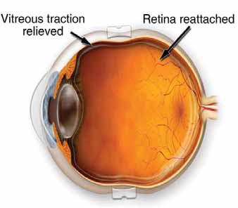
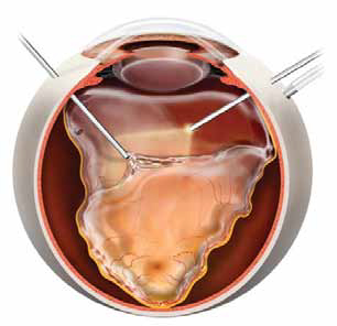
2. Vitrectomy: Tiny incisions are made in the sclera (white of the eye), and small instruments are placed into the eye to reattach the retina. A gas bubble is then injected into the eye to place pressure on the retina to help keep it attached. In addition, a laser is applied to the retina to hold the retina in place. During the healing process, the natural fluid of the eye gradually replaces the gas bubble and refills the eye. This surgery is usually performed in an operating room under local anesthesia with mild sedation.
3. Scleral buckle: A tiny synthetic band is attached to the outside of the eyeball to gently push the wall of the eye against the detached retina. During this procedure, cryotherapy or laser is also used to help hold the retina in place. This surgery is usually performed in an operating room under local anesthesia with sedation or general anesthesia.
4. Pneumatic retinopexy: A small gas bubble is injected into the eye that pushes the retina back against the wall of the eye. Laser or cryotherapy is then used to seal the retinal tear and help hold the retina in place. This surgery is performed in an office setting under topical or local anesthesia. Recovery time can be much quicker with pneumatic retinopexy. However, not all patients will be good candidates for this technique.
